

Museum Veterinary Services Team comes to aid of resident balloonfish
For immediate release ‐ March 12, 2021
Contact: Micah Beasley, 919.707.9970. Images available upon request
The balloonfish, a Long-spine Porcupinefish (Diodon holocanthus), is a marine species native to North Carolina. Their teeth are fused together into a sharp cranial beak-like structure ideal for crushing prey such as clams, sea urchins and crabs.
Although balloonfish are generally slow swimmers, they have a well-developed defensive behavior used to escape predators. When stressed or challenged, they gulp water (or air if held out of the water) and inflate their bodies into a spiny ball up to four times the volume of the fish in a relaxed state.
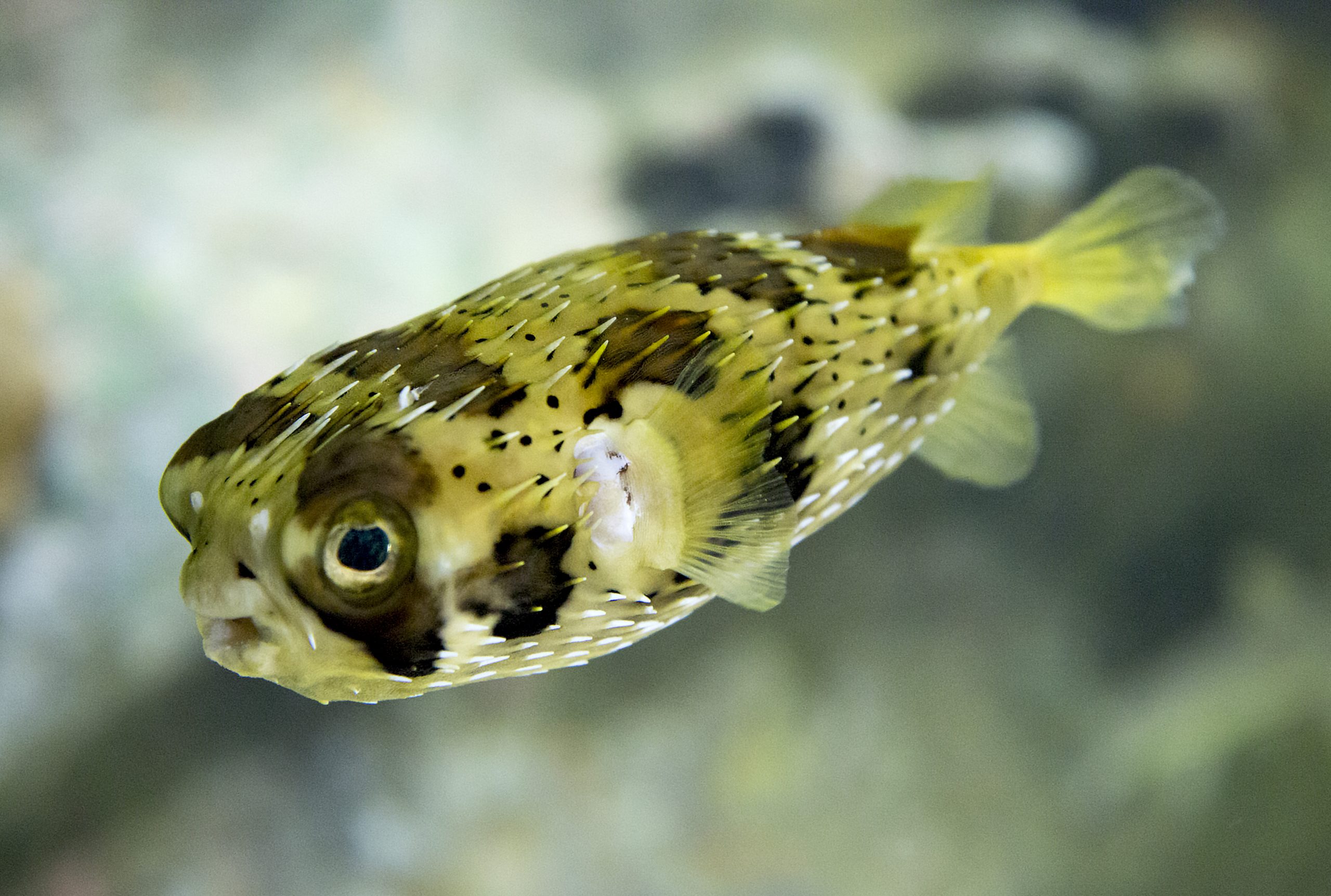 The Museum’s resident balloonfish as a young adult in its relaxed state. Photo: Karen Swain/NCMNS.
The Museum’s downtown location has a resident balloonfish (pictured above; click to enlarge) who is popular with guests and staff alike.
However, after presenting to the Veterinary Services team with a recent history of lethargy, anorexia and potential abnormal buoyancy (struggling to swim to the surface), the team had to spring into action to see what was ailing this marine marvel.
A medical examination revealed a firm mass within the body cavity of the fish. Next, it was critical for the team to determine if the mass was causing blockage of the GI tract.
The team also needed to evaluate the fish’s swim bladder (an internal air sac that aids with buoyancy).
Use of imaging tools and techniques are valuable in cases like this to examine a creature’s internal structure, allowing the team to visualize normal and abnormal structures that would otherwise go unseen.
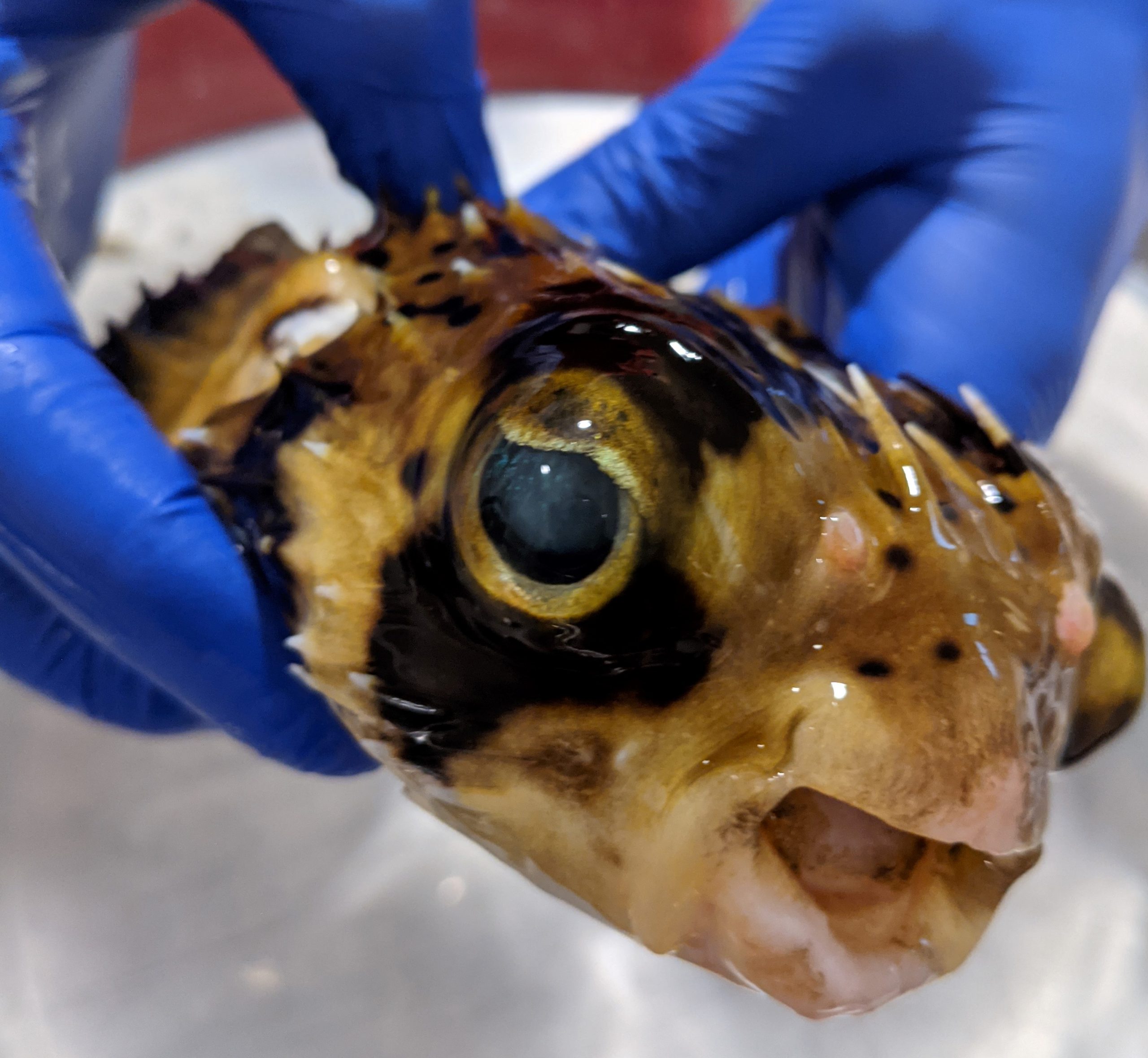 The Museum’s balloonfish under anesthesia. Click to enlarge.
In preparation for medical examinations, or to facilitate medical procedures such as a computer tomography (CT) scan, fish can be anesthetized and removed from water for short periods of time.
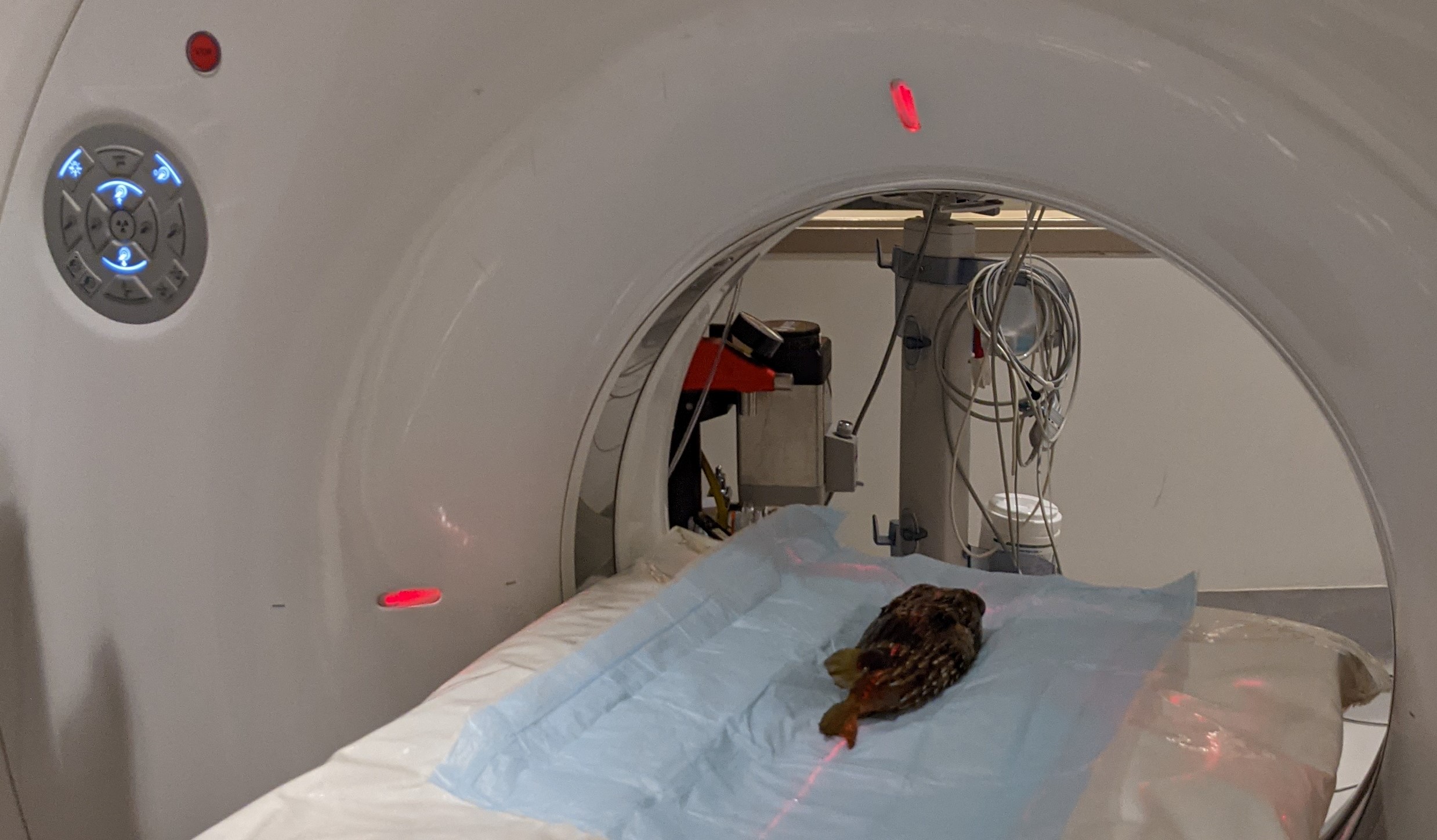 The Museum’s balloonfish getting his CT scan. Click to enlarge.
CT is a common modality used to visualize details of internal organs, especially contrasting air, soft tissue and mineralized structures, such as bone or shell. Additionally, colors and textures can be applied to contrast and differentiate tissues and material types in image reconstructions.
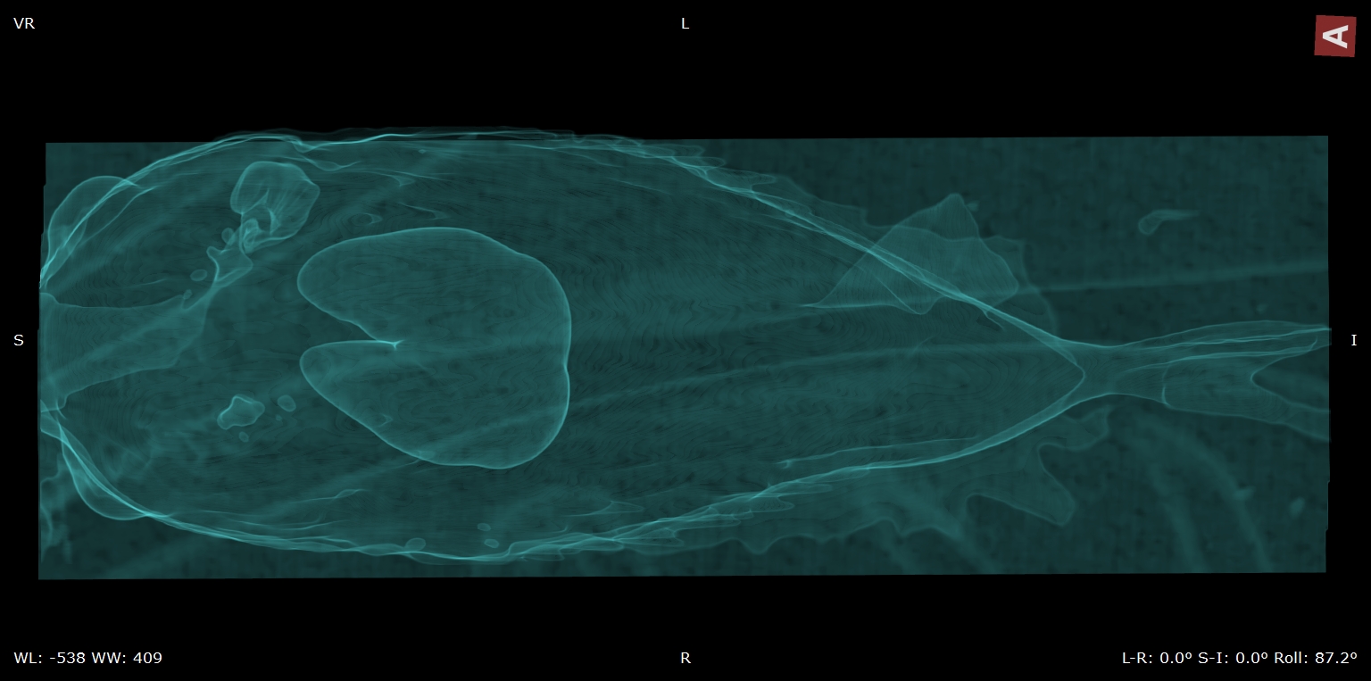
This CT scan image of the balloonfish (above; click image to enlarge) provides contrast to aid in the evaluation of gas within the fish, causing it to stand out visually from internal tissues and structures. The U-shaped structure highlighted in the center of the image is the gas-filled swim bladder. This is a normal feature for this species.
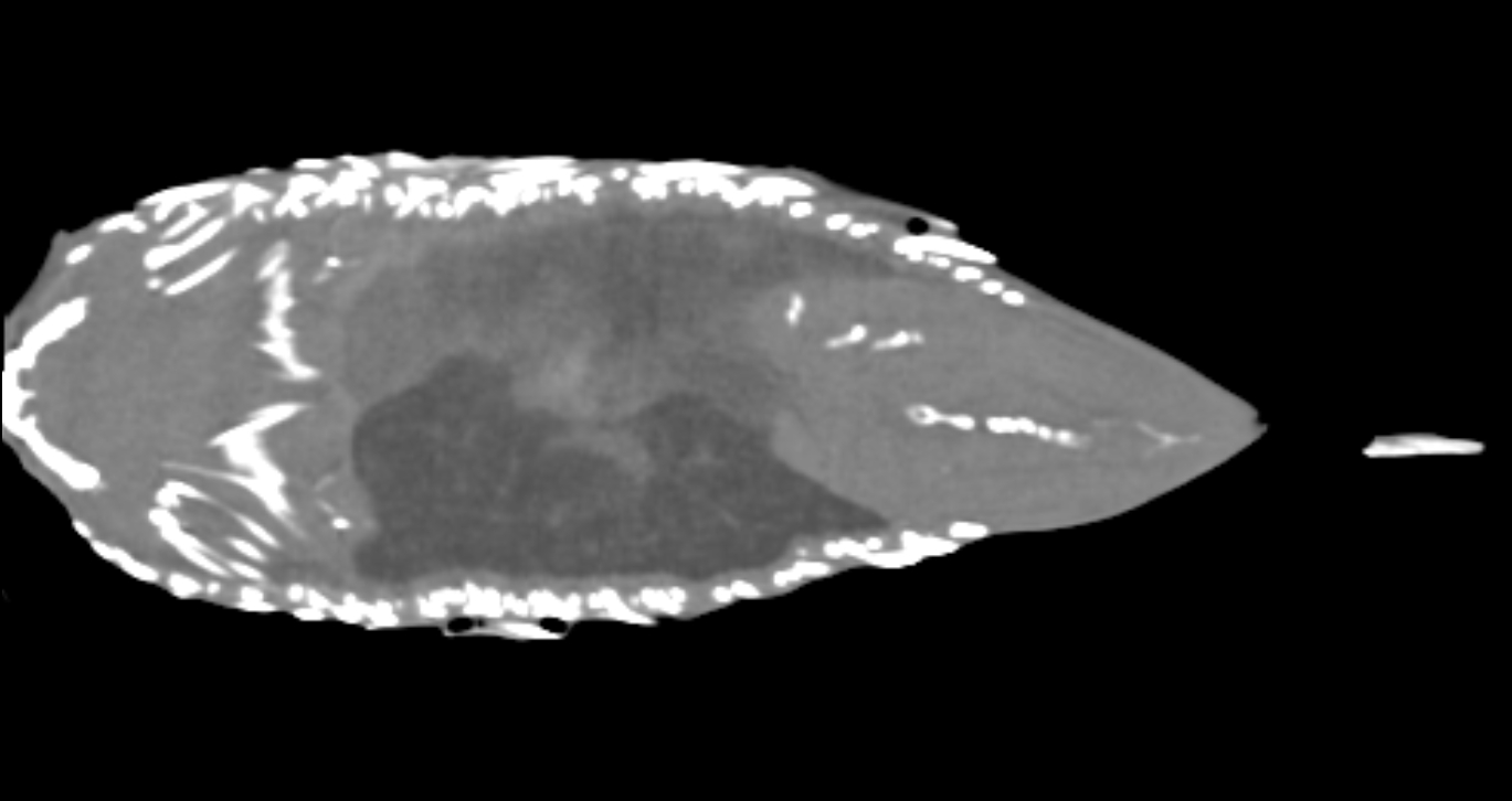
This second CT scan image (above; click image to enlarge) demonstrates a single approximate frontal plane view contrasting bone (white) from other tissues, organs, and structures (shades of gray). Based on the full reconstruction from this scan, the team saw evidence of liver disease and some abnormal calcification within the left kidney. However, the GI tract appeared to be clear with no evidence supporting an abnormal mass within the body cavity.
After further evaluation, the Veterinary Services team suspects that the reported mass was within the GI tract, however, the medical exams and handling of the fish helped it to pass the mass. The team is pleased to report that the balloonfish has returned to good health.
The imaging data captured by the team will serve as a baseline for future medical evaluations.
Bonus video: the Veterinary Services team captured incredible, 3D color imaging that demonstrates the extensive bony dermal structures (spines) in the skin of this species.