

Off with its head!
For immediate release ‐ May 28, 2024
Paleontology
Contact: Jon Pishney, 919.244.7913. Images available upon request
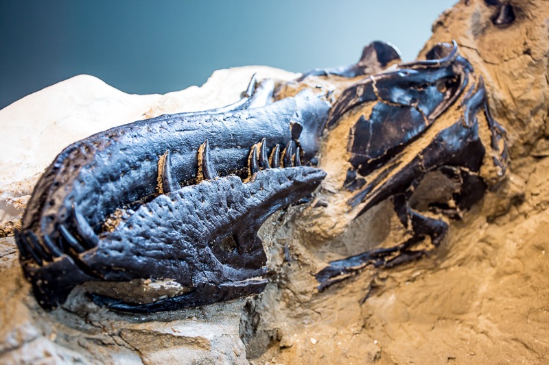 The Dueling Dinosaurs tyrannosaur skull. Photo: NCDNCR.
Just like your skull, the Dueling Dinosaurs tyrannosaur’s skull once housed and protected essential soft tissues such as the brain, the inner ear and sinuses. Studying these tissues can help us understand the senses and physiology of our predator, but they decomposed long ago during the fossilization process.
Luckily, we have ways around this problem. By using a medical technology known as microCT (X-ray computed tomography), we can create detailed, 3D scans of the inside of the skull without damaging it, including places we can’t see with the naked eye. This technology allows us to reconstruct the shape of organs, nerves, blood vessels and other hidden structures by digitally filling the voids in the fossilized bones that once surrounded them.
Once removed, our tyrannosaur skull was protected with a plaster casing, cradled in foam and carefully driven to the University of Texas High Resolution X-ray CT Facility in Austin by Dueling Dinosaurs project postdoctoral researcher Dr. James Napoli. There it underwent a two-day long, high-radiation scan, necessary to penetrate the dense rock and fossil bone.
Can you imagine sitting still in a CT machine for two days with a broken leg? Be grateful you aren’t a tyrannosaur.
For more information about our upcoming activities, conservation news and groundbreaking research, follow @NaturalSciences on Instagram and Facebook.